|
|
Lacunar Skull
Luckenschadel Skull
General Considerations
- Bone dysplasia of skull consisting of multiple oval lucencies separated by dense, bony ridges
- Associated with
- Neural tube defects, especially myelomeningocele
- Chiari II malformation
- Encephalocele
- Not related to degree of concomitant hydrocephalus
- Inner table more affected than outer
Clinical Findings
- Present at birth
- Unrelated to increased intracranial pressure
Imaging Findings
- Well-defined lucent areas in calvarium representing nonossified fibrous bone
- Lacunae are bounded by normally ossified bone
- Most prominent in parietal bones
- Small posterior fossa associated with Chiari II malformation
Differential Diagnosis
- Normal convolutional markings seen during a period of rapid brain growth (3 to 7 years).
- Increased convolutional markings associated with synostosis at an older age
- Increased intracranial pressure - beaten-silver appearance; closed sutures; may have abnormal skull size
Treatment
- Appearance resolves spontaneously by age 6 months
Prognosis
- Spontaneously disappears by 4-6 months old

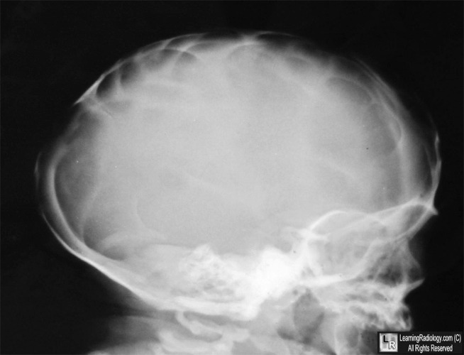
Lacunar Skull. There are multiple focal areas of radiolucency in the skull (white arrows)
bounded by more normal, dense bony ridges. The child had a known myelomeningocele.
For this same photo without the arrows, click here
For more information, click on the link if you see this icon 
The Infant Skull: A Vault of Information. RB J Glass, S K Fernbach, KI Norton, PS Choi and TP Naidich. March 2004 RadioGraphics, 24, 507-522.
|
|
|
